
Basic Cell Culture Workflow: Prepare, Grow & Analyze
If you have never worked with cells before, this is the right place for you. We will help you understand the basics of cell culture in this blog. Those who are experts in cell culture, you might find this useful. If not, please share your experience in the comment section below and help make this blog better.
Basically, the cell culture workflow includes preparation, growth and analysis. Let us look into the different stages in details.
Preparation Phase of Cell Culture
Cells
As the foundation of cell culture, cells are the essential element. Our focus on cell culture would be incomplete without cells. Hence, to make your life easier in the cell culture journey, we provide a wide variety of cells lines from Sigma-Aldrich, which partnered with the European Collection of Authenticated Cell Cultures (ECACC). It has more than thousands of cell lines available, and all of them are high in purity and quality. Not to say that they are low in passage number as well.
Culture Media Sterilization
If the volume of culture media is small, you can filter-sterilize it using a syringe. However, if you need to filter-sterilize large amounts of culture media, using a syringe filter costs much more time and money.
So, how can you filter-sterilize a large amount of media efficiently? One of the ways is to filter-sterilize your culture media with Stericup® Quick Release. This filter uses a 0.22m pore size, fast-flow, low protein binding membrane. The fast flow is crucial. If the media expose to air for too long, the pH might change and affects the pH-sensitive cells.
During the preparation phase, you must be extremely cautious not to contaminate your culture with bacteria, or worse, mycoplasma. As mycoplasma are too small to be observed under the microscope, you need to use the LookOut® Mycoplasma PCR detection kit. This kit uses a polymerase chain reaction to detect Mycoplasma, Acholeplasma, and Ureaplasma contamination in cell cultures.
Our recommendation for storing your tissue or cells is Cryostor®. It is a safe and protective environment for your tissues and cells while being frozen, thawed and stored.
Growth Phase of Cell Culture
It is necessary to grow and feed your cells with suitable cell culture media, including Dulbecco Modified Eagle Medium (DMEM), Minimum Essential Medium Eagle (EMEM), Fetal Bovine Serum (FBS), etc. Our recommendation is to use the right culture media for the cells.
Liquid Handling Tools
Now that you have cells and culture media, it is time to grow your cells. You can culture your cells on the cell culture flasks or cell culture plates. In some cases, you may need a cell adhesion reagent, such as laminin. Laminin helps cells attach nicely to the surface. Lastly, you would need some liquid handling tools, such as Eppendorf® micropipettes, centrifuge tubes and cell strainer.
Analysis phase of Cell Culture
Immunoassay
In some cases, after growing your cells, you may need to analyze them using the immunoassay method. These methods typically include ELISA, Western blotting, immunofluorescence, immunohistochemistry, flow cytometry, etc. These immunoassays require antibodies, and Zoomab® Recombinant Antibody is an excellent choice.
As the new generation of recombinant monoclonal antibodies, Zoomab® Recombinant Antibody offers superior specificity and reproducibility. The reason is that recombinant antibodies were created by cloning antibody genes into expression vectors without the use of hybridomas. As a result, any problem associated with hybridomas, (e.g. cell line drift, antibody expression variation and antibody sequence mutation) will not occur. On top of that, it reduced the use of animals in the research and provided an excellent batch-to-batch consistency.
Cell Count
Other than immunoassay, you might want to calculate the number of cells in your culture. Traditionally, you can count cells manually with a hemacytometer. Using a haemacytometer indeed is the easiest and the most common way to count the number of cells. However, the results are often inconsistent due to the bias of the users, not to mention that the process is also time-consuming.
To avoid these problems, why not use an automated cell counter, Scepter⢠3.0? Scepter⢠3.0 is 7 to 10 times faster than the hemacytometer. It is also faster than most automated cell counters. In addition, Scepter⢠3.0 count the cells using the principle of Ohm’s law. The voltage changes as cells pass through the sensor. ScepterTM 3.0 records each change in voltage as spikes. ScepterTM 3.0 presents data as a histogram, and you can determine the concentration and number of cells from the histogram. On top of that, the data collected is from thousands of cells per sample, resulting in more precise counts.
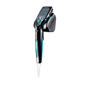
Automated Handheld Scepter⢠3.0
Conclusion
In short, the basic cell culture workflow includes prepare, grow and analyze. If you want to know more about other topics, please let us know in the comment section below. We will do our best to post new information blogs.


