Showing 1–24 of 29 results

Show sidebar

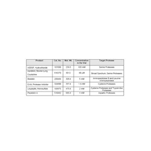
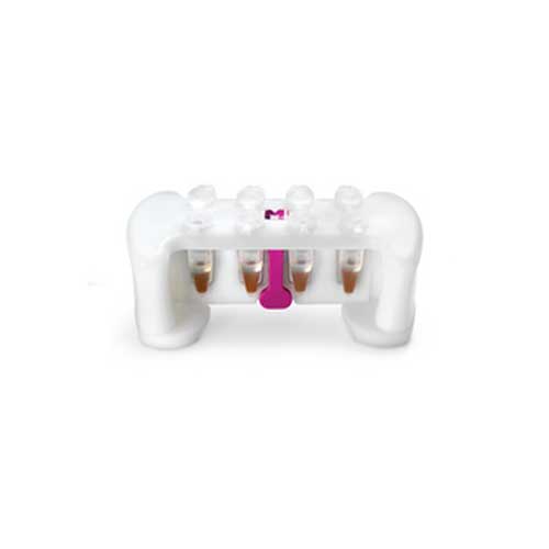
Affinity purification tools

Brand:

Merck

Description

Merck Millipore offers a wide range of tools for protein purification, including affinity magnetic beads and affinity agarose resins.
• PureProteome™ magnetic beads are ideal for small volume affinity purification assays, such as
immunoprecipitation and serum depletion or enrichment.
• Affinity agarose portfolio for larger volume applications, such as antibody purification and recombinant
protein purification

Amicon® Ultra Filter

Brand:

Merck

Description

Applications Include:
Concentration of biological samples containing antigens, antibodies, enzymes, nucleic acids, or microorganisms.
Purification of macromolecular components found in tissue culture extracts or cell lysates.
Primer removal, PCR cleanup, removal of linkers or macromolecular labels from a reaction mix, and protein removal prior to HPLC.
Desalting, buffer exchange and protein dialysis.

Anti-acetyl-Histone H3 (rabbit polyclonal)

Brand:

Merck

Description

Histone H3 is one of the five main histone proteins involved in the structure of chromatin in eukaryotic cells. Featuring a main globular domain and a long N-terminal tail, H3 is involved with the structure of the nucleosomes of the ‘beads on a string’ structure. The N-terminal tail of histone H3 protrudes from the globular nucleosome core and can undergo several different types of epigenetic modifications that influence cellular processes. These modifications include the covalent attachment of methyl or acetyl groups to lysine and arginine amino acids and the phosphorylation of serine or threonine. Acetylation of histone H3 occurs at several different lysine positions in the histone tail and is performed by a family of enzymes known as Histone Acetyl Transferases (HATs). Acetylation of lysine14 is commonly seen in genes that are being actively transcribed into RNA. Research focus area: Epigenetics.

Key Applications:
-Western Blotting
-Immunocytochemistry
-ChIP-seq
-Chromatin Immunoprecipitation (ChIP)

Anti-Actin, clone C4 (mouse monoclonal)

Brand:

Merck

Description

Actin is an abundant cytoskeletal protein found in all cells (True, 1990). The protein’s 42 kD peptide chain assumes two physical forms: globular actin, which may serve as a cytoplasmic storage pool, and fibrous actin, which, in conjunction with myosin, generates muscle contraction (True, 1990). In non-muscle cells, actin appears to be involved in a variety of functions, such as cell motility, exocytosis, and phagocytosis (True, 1990). Distribution of the six known isoforms of actin – four muscle actins (alpha-skeletal, alpha-vascular smooth, alpha-cardiac, and gamma-enteric smooth) and two cytoplasmic actins (alpha and gamma) is tissue specific (Otey, 1986; Lessard, 1988). Research focus area: Cell Structure

Key Application:
-ELISA
-Immunocytochemistry
-Immunofluorescence
-Immunohistochemistry
-Immunohistochemistry (Paraffin)
-Western Blotting

Anti-Glutamate Receptor 2, extracellular, clone 6C4, Alexa Fluor® 488 Conjugate

Brand:

Merck

Description

Glutamate receptors (GluRs) can be categorized as ionotropic or metabotropic and subcatergorized by their agonist preferences (NMDA, AMPA or Kainic acid). There are four types of AMPA selective GluR subunits (GluR1, GluR2, GluR3 and GluR4). Tetrameric or pentameric combinations of different subunits contributes to the functional diversity of AMPA receptors. In general, AMPA receptors mediate fast synaptic current at most excitatory synapses, with stoichiometry characterized by subtype composition. Although subunit composition of AMPA receptors varies, they must contain at least one edited GluR2 subunit to be calcium impermeable. The critical residue controlling calcium permeability is in the pore loop region. In GluR1, GluR3, and GluR4, this positionis occupied by a Gln residue. In GluR2, it is occupied by an Arg residue. It has been shown experimentally that the presence of Arg in this position blocks CA2+ ion permeability, while a Gln does not. Relative calcium permeability in AMPA receptor channels may be significant in pathological neurotoxic damage and long term changes in nervous system responses. Research focus area: Neuroscience

Key Applications:
-ELISA
-Immunocytochemistry
-Immunohistochemistry
-Immunoprecipitation
-Radioimmunoassay
-Western Blotting

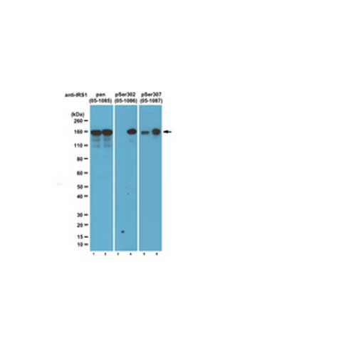
Anti-IRS1, clone 4.2.2 (mouse monoclonal)

Brand:

Merck

Description

IRS1 (Insulin Receptor Substrate 1) transmits insulin signals via metabolic and mitogenic pathways. IRS1 is heavily phosphorylated on both serine and tyrosine residues. These phosphorylated tyrosines enable IRS to act as a docking protein that binds SH2 domains of such proteins as PI3 Kinase (phosphatidylinositol 3-kinase) and GRB2, resulting in activation. Over expression and phosphorylation of serine is associated with insulin resistance and breast cancer. Some of the more notable phosphorylation sites are Ser302 that is phosphorylated following insulin stimulation. Ser307, phosphorylated by JNK and IKK, is a key regulatory site that appears to disrupt the IRS1/IR interaction and inhibits insulin-mediated activation of the PI3 kinase and MAPK pathways, and Ser636/639 that is known to be phosphorylated by p70S6K downstream of mTOR and acts as a negative feedback loop. Research focus area: Signaling

Key Applications:
-Western Blotting
-Immunoprecipitation
-Immunocytochemistry

Anti-p62 (Sequestosome-1), clone 11C9.2 (mouse monoclonal)

Brand:

Merck

Description

p62 is also known as sequestosome-1, and is a 62 kDa ras-GAP associated protein. p62 is thought to bind to src-family tyrosine kinases and may also provide a linker action for activated src-family kinases with their downstream effectors. p62 has an RNA-binding region and may have RNA-binding ability. p62 is associated with activation of transcription factor NF-κB, involved in cell differentiation, immune response and potassium channel regulation. p62 has recently been linked to extrinsic apoptosis and may be a key player in tumorigenesis. Research focus area: Cancer.

Key Applications:
-Western Blotting
-Flow Cytometry
-Immunocytochemistry

Anti-phospho Histone H2A.X (Ser139), clone JBW301, Alexa Fluor® 488 Conjugate

Brand:

Merck

Description

Histone H2A.X is a variant of histone H2A, and is similarly associated with genomic DNA. However, histone is structurally different from other members of the H2A family in possessing a C-terminal tail that contains the Ser139 residue that is phosphorylated in response to breaks in double-stranded DNA. The phosphorylation of H2A.X is a rapid process that is mediated by ATM/ATR proteins.

Anti-phospho Histone H2A.X (Ser139), clone JBW301, Alexa Fluor® 647 Conjugate

Brand:

Merck

Description

As a member of the histone H2A family, histone H2A.x (H2A.x) is a variant histone H2A which replaces conventional H2A in a subset of nucleosomes. H2A.x is involved in the DNA repair of double-strand breakage (DSB) damage on nuclear DNA. After a double strand DNA break, H2A.x is rapidly phosphorylated at serine 139 by ATM kinase and becomes gamma-H2AFX. This phosphorylation step can extend up to several thousand nucleosomes from the actual site of the DSB and may mark the surrounding chromatin for recruitment of proteins required for DNA damage signaling and repair. As a part of posttranslational modifications during apoptosis caused by severe DNA damage, high expression of phosphorylated H2A.x is considered as an accurate indicator of apoptosis.

Anti-phospho-Histone H2A.X (Ser139), clone JBW301 (mouse monoclonal)

Brand:

Merck

Description

Histone H2A is one of the 5 main histone proteins involved in the structure of chromatin in eukaryotic cells. Featuring a main globular domain and a long N terminal tail H2A is involved with the structure of the nucleosomes of the ‘beads on a string’ structure

Anti-phospho-MYPT1 (Thr696) Antibody – Discontinued

Brand:

Merck

** This product has been discontinued, please contact us for more information.

Description

Myosin phosphatase target subunit 1 (MYPT1), also called Myosin-binding subunit of Myosin phosphatase, is one of the subunits and an integral component of the Myosin phosphatase. Myosin phosphatase regulates the interaction of Actin and Myosin downstream of the guanosine triphosphatase Rho, which inhibits Myosin phosphatase through the action of Rho-kinase. It is also involved in smooth muscle contraction. Inhibition of Myosin light chain phosphatase results in Ca2+ sensitization of smooth muscle contraction. This inhibition is modulated through phosphorylation of MYPT1 by a ZIP-like kinase, which associates with MYPTI and phosphorylates the inhibitory site in smooth muscle. The phosphorylation of MYPT1 by protein kinase C results in altered dephosphoryation of Myosin by attenuating the binding of protein phosphatase 1 catalytic subunit (PP1c) and the phosphorylated 20 kDa Myosin light chain to MYPT1. PP1c interacts with at least four binding sites on the amino-terminus of MYPT1. MYPT1 is localized on stress fibers, and is distributed close to the cell membrane and at cell-cell contacts to regulate Myosin phosphatase activity.

BugBuster® Protein Extraction Reagent

Brand:

Merck

Description

Protein stability is fundamental to all aspects of protein research, including analysis by Western blotting. Combine our gentle protein extraction kits with protease inhibitors to obtain stabilized, intact and active proteins.

BugBuster® Master Mix combines BugBuster Protein Extraction Reagent with Benzonase®Nuclease and rLysozyme™ Solution in one convenient reagent. BugBuster Master Mix allows for maximum recovery of active soluble protein from both Gram-negative and Gram-positive bacteria. With the Master Mix, there is no need for dilution or separate addition steps. The two available package sizes provide sufficient reagents for protein extraction from 20 g and 100 g cell paste.

Chymostatin (Microbial Streptomyces), 100 mg

Brand:

Merck

Description

Chymostatin. M.W. 604.92
Mixture of types A, B and C. Inhibits Chymotrypsin-like serine proteases including alpha, beta, gamma and delta chymotrtrypsins.
Sequence:
[(S)-1-Carboxy-2-Phenylethyl]-Carbamoyl-a-[2-Iminohexahydro-4(S)-Pyrimidyl]-(S)-Glycyl-X-Phenylalaninal
X: L-Leu (type A), L-Ile (type B), L-Val (type C)
Working concentrations 10-100μM.
5mg/825 microliters in DMSO = 10mM solution.
Dilute 1:100 for 100μM working solution.

Lyophilized. Dissolve in dimethylsulfoxide (DMSO). After powder is thoroughly dissolved, dilute the DMSO solution with 0.9% NaCl. The concentration of DMSO should be maintained at 10% or higher. Otherwise chymostatin may precipitate from the solution.

Maintain lyophilized material at -20ºC for up to one year. Store reconstituted product in aliquots at -20ºC for up to 3-4 weeks. Do not thaw and refreeze.

CytoBuster® Protein Extraction Reagent (for mammalian cell lysis)

Brand:

Merck

Description

The CytoBuster™ Protein Extraction Reagent is a proprietary formulation of detergents optimized for efficient extraction of soluble proteins from mammalian and insect cells. The gentle, non-ionic composition of CytoBuster Protein Extraction Reagent enables isolation of functionally active endogenous or expressed proteins without a need for secondary treatment such as sonication or freeze/thaw. CytoBuster Protein Extraction Reagent has been specifically formulated for utilization in Western blotting, immunoprecipitation, and kinase/phosphatase assays. The reagent is compatible with protease inhibitors, kinase inhibitors, and phosphatase inhibitors.

D-Tube™ Dialyzer Mini, Midi, Maxi and Mega

Brand:

Merck

Description

The D-Tube™ Dialyzers be used for dialysis and electroelution of proteins, RNA, DNA, and oligonucleotides from polyacrylamide or agarose gels. The disposable, single-use tubes require no syringes, microcentrifuge, or laborious steps to manipulate small sample volumes. The sample is added and removed using a standard laboratory pipet. Available with molecular weight cutoffs (MWCO) from 3.5 to 14 kDa, the D-Tube Dialyzers are designed in four volume capacities: mini (10-250 µl), midi (50-800 micro;l), maxi (100-3000 µl), and mega (10ml, 15ml, and 20 ml). The membrane is ultra-clean, EDTA-treated regenerated cellulose, sulfur- and heavy metal-free.

The D-Tube Dialyzers Mini, Midi, and Maxi contain 10 D-Tube Dialyzers and one floating rack that can hold up to four D-Tube Dialyzers. The D-Tube Dialyzers Mega contain 10 or 50 tubes and one or 5 floating racks to hold one D-Tube Mega in the exchange buffer.

Goat anti-Mouse, FITC conjugate

Brand:

Merck

** This product has been discontinued.

Description

Immunoglobulin G (IgG) is a multimeric immunoglobulin, built of two heavy chains γ and two light chains. Each complex has two antigen binding sites. It is one of the most abundant proteins in human serum with normal levels between 8-17 mg/ml in adult blood. IgG is important for our defense against microorganisms and the molecules are produced by B lymphocytes as a part of our adaptive immune response. The IgG molecule has two separate functions; to bind to the pathogen that elicited the response and to recruit other cells and molecules to destroy the antigen. Furthermore, this is the only isotype that can pass through the human placenta, thereby providing protection to the fetus in its first weeks of life before its own immune system has developed. The variability of the IgG pool is generated by somatic recombination and the number of specificities in an individual at a given time point is estimated to be 1011 variants.

Immobilon-P PVDF Membrane, 0.45 μm

Brand:

Merck

Description

Immobilon-P Transfer Membrane:

  • 45 µm pore size Recommended for most western blotting, especially proteins greater than 20 kDa
  • Millipore’s Rapid Immunodetection Protocol reduces detection times by up to 2 hours by eliminating membrane blocking and several wash steps with no loss of sensitivity or specificity

Features & Benefits:

  • Won’t crack, curl or fracture when cut
  • Low background
  • Superior staining capabilities
  • Can be reprobed multiple times

Applications:

  • Western Blotting, Immunoblotting, Dot Blotting, Protein Sequencing;
  • Compatible with Chemiluminescent, Fluorescent, Radioactive, Chromogenic, and Chemifluorescent Detection
  • Compatible with all commonly used fluorescent probes at all excitation and emission wavelengths

Immobilon® Block

Brand:

Merck

Description

Immobilon® Block Noise Cancelling Reagents are designed especially for Western blotting applications. This family of protein-free, ready-to-use buffers has been optimized to reduce background levels when using chemiluminescence, fluorescent, or phosphotyrosine detection. Bløk buffers are ideal for use with Immobilon®-P Western blotting membranes and the SNAP i.d.® system, and are also compatible with standard Western blotting techniques.

Immobilon®-FL Membrane, 0.45 μm

Brand:

Merck

Description

Immobilon-FL Membrane:
– The first transfer membrane optimized for fluorescence applications
– Extremely low background improves sensitivity of all fluorescence detection protocols
– Compatible with all commonly used fluorescent probes at all excitation and emission wavelengths
– Ideal for multiplexing and chemifluorescence applications

Features & Benefits:
– Won’t crack, curl or fracture when cut
– Low background
– Superior staining capabilities
– Can be reprobed multiple times

Applications:
– Western Blotting, Immunoblotting, Dot Blotting, Protein Sequencing;
– Compatible with Chemiluminescent, Fluorescent, Radioactive, Chromogenic, and Chemifluorescent Detection
– Compatible with all commonly used fluorescent probes at all excitation and emission wavelengths

Immobilon®-PSQ PVDF Transfer Membrane, 0.2 μm

Brand:

Merck

Description

Immobilon-PSQ Membrane:
– 0.2 µm pore size and a large internal structure
– Higher protein adsorption and sequencing yields than other membranes
– Recommended for blotting proteins less than 20 kDa
– Prevents blow-through of low molecular weight proteins

Features & Benefits:
– Won’t crack, curl or fracture when cut
– Low background
– Superior staining capabilities
– Can be reprobed multiple times

Applications:
– Western Blotting, Immunoblotting, Dot Blotting, Protein Sequencing;
– Compatible with Chemiluminescent, Fluorescent, Radioactive, Chromogenic, and Chemifluorescent Detection
– Compatible with all commonly used fluorescent probes at all excitation and emission wavelengths

Immobilon™ Western HRP Substrate

Brand:

Merck

Description

Chemiluminescent HRP* substrates (also known as ECL reagents) are the most sensitive reagents used in the detection of Western blots. The Immobilon™ Western HRP Substrates are a family of three premixed HRP substrates, which offer several advantages over other detection reagents.

Key Application:
Western Blotting

Leupeptin, 100 mg

Brand:

Merck

Description

Leupeptin is an organic compound produced by actinomycetes, serine and cysteine proteases reversibly. Inhibits trypsin-like proteases and some cysteine proteases including endoproteinase Lys-C, Kallikrein, papain, thrombin, cathepsin B and trypsin.
MW:426.56 s 49.04 . 18.02 {C20H36N6O4 . ½H2SO4 . H2O}
Ac-Leu-Leu-Argininal (hemi-sulfated form). Thus material has three tautomeric isomers that can be visualized by chromatography.
Appearance: White amorphous powder
Working range 10-100μM. Stable for several hours in water. Stock solutions of 10mM in water are stable for 1 week at 4ºC and 6 months at -20ºC.
5mg/1.054 mL = 10mM. Typically dilute 1:100 for a 100μM working solution.
Source: Organic isolated from actinomycetes.

Maintain dry at -20ºC for up to 18 months after date of receipt. Store reconstituted product in aliquots at -20ºC for up to 6 months. Avoid repeated thaw freeze cycles.

ProteoExtract® Native Membrane Protein Extraction Kit

Brand:

Merck

Description

A convenient kit for the isolation of native integral membrane and membrane-associated proteins, without the need for ultracentrifugation. Extraction is based on association of proteins with cellular membranes rather than on their hydrophobicity. Resulting samples are suitable for use in functional assays, 2-D gel electrophoresis, and other applications.

Note: 1 kit is sufficient for up to 20 sample extractions.

ProteoExtract® Subcellular Proteome Extraction Kit

Brand:

Merck

Description

Fast and reproducible extraction of subcellular proteomes from mammalian cells

ProteoExtract® Subcellular Proteome Extraction Kit (S-PEK) is designed for fast and reproducible extraction of subcellular proteomes from adherent and suspension-grown mammalian cells. The S-PEK takes advantage of the different solubilities of certain subcellular compartments in the four selected reagents. In the case of adherent cells, the procedure is performed directly in the tissue culture dish without the need for cell removal. Cells or the parts of the cells remain attached to the plate during sequential extraction of subcellular compartments, until the appropriate extraction reagent is used. Thus, the early destruction of the cellular structure by enzymatic or mechanical detachment of cells from the tissue culture plate and any mixing of different subcellular compartments is prevented. For suspension-grown cells, extraction starts with gentle sedimentation and washing of the cells. The stepwise extraction delivers four distinct protein fractions from one sample:

– Cytosolic fraction (F1)
– Membrane/organelle protein fraction (F2)
– Nucleic protein fraction (F3)
– Cytoskeletal fraction (F4)

Proteins are obtained in the native state making the S-PEK suitable for many downstream applications such as 1D and 2D gel electrophoresis, immunoblotting, enzyme activity assays, and protein microarrays.

Sample size: 3-5×10⁶ or 25-50 mg tissue.